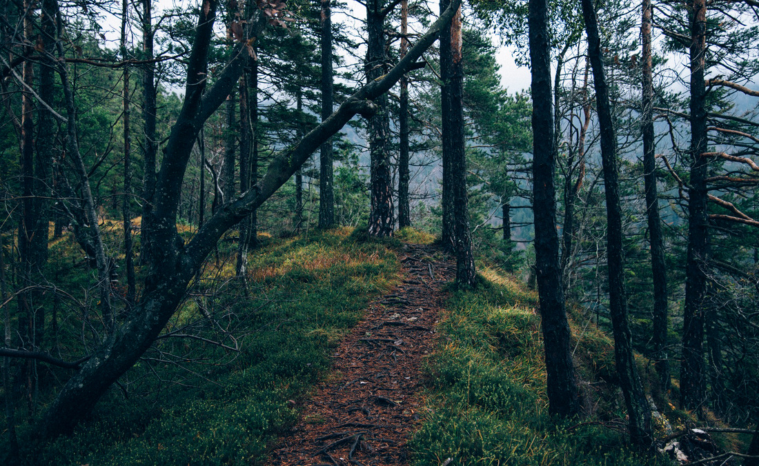
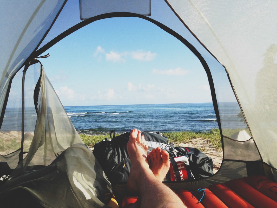
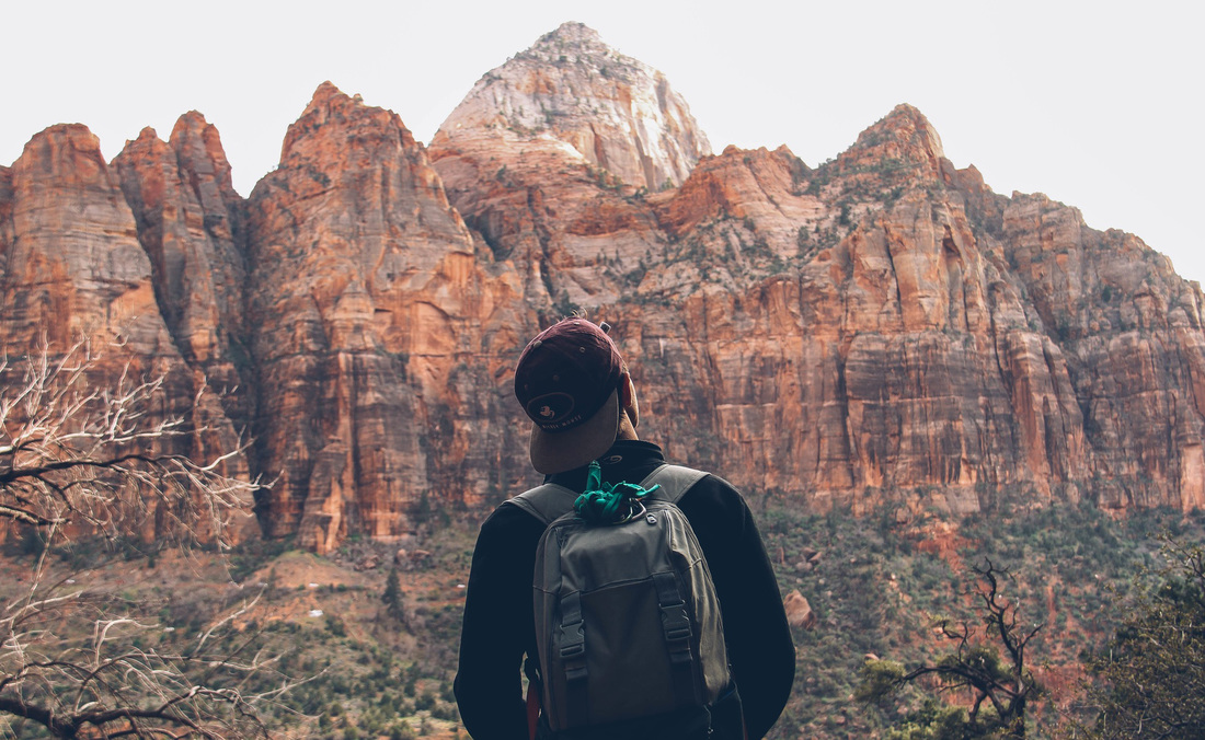
I'm James. This is my year of travel.

Weissfeld, 12th ed 2007, Publisher Elsevier. Bailey & Scott’s Diagnostic Microbiology.Simmous, 4th ed, Publisher Churchill Living Stone, New York, Melborne, Sans Franscisco 1996. Mackie and Mc Cartney Practical Medical Microbiology.Lehman & George Manuselis, 3rd edition2007, Publisher Elsevier. Collier, 8th ed 1990, Publisher Edward Arnold publication, London. Roberts, 3rd ed 1985, Publisher Williams and Wilkins, Baltimore. Isenberg, Albert Einstein College of Medicine, New York, Publisher ASM (American Society for Microbiology), Washington DC. Clinical Microbiology Procedure Handbook Vol.3 rd ed 1988 Publisher WB Saunder co, Philadelphia. The pathogenic fungi and the Pathogenic Actinomycetes. Editors: Emmons and Binford, 2nd ed 1970, Publisher Lea and Febiger, Philadelphia. Other Information related to CryptococcusĬryptococcus capsule in Giemsa stained smear as shown below-Ĭryptococcus neoformans growth on Sabouraud Dextrose Agar (SDA) Bird Seed Agar (BSA)- Niger seed is replaced by Nescafe Coffee and Urea Hydrolization Test-Positive as shown in Video-Ĭryptococcus neoformans in LPCB tease mount as shown below. Some strains of Cryptococcus neoformans , as well as other cryptococci, may not produce discernible capsules in vitro.Leukocytes and tissue cells may be dissolved by adding a drop of 10% KOH. Fat droplets, white blood cells, and tissue cells are sometimes confused with organisms like Cryptococcus neoformans cells.India ink preparation is only for presumptive identifications of organisms and therefore it needs other tests like biochemical, immunological, molecular, or mass spectrometry testing that must be performed on colonies from pure culture for complete identification.Although this is only a small change, it can dramatically change the end result when. The main difference is that India ink only uses soot and water where as Sumi ink uses soot and animal glue (usually gelatin in the more modern formulas). Limitations of Ink Ink or Negative staining As we touched on above, the ingredients for Sumi ink is slightly different to the ingredients of India ink. Do not make the preparation too thick otherwise, the cells and capsules will not be seen.When India ink is not available, use the nigrosin (20% w/v) solution.Pelikan black drawing ink is suitable for this test.a presumptive diagnosis of cryptococcal meningitis can be made. When encapsulated yeasts are detected in C.S.F. Negative control: Absence of encapsulated yeasts seenĬapsules of Cryptococcus neoformans like organisms seen Importance of this test.Positive control: Presence of encapsulated yeasts seen.Look for oval or round cells, some showing budding, irregular in size, measuring 2-10 µm in diameter, and surrounded by a large unstained capsule as shown in the above figure. Now, examine the preparation under a microscope using the 40 X objective.a drop of the sediment to a slide and add a drop of India ink. Transfer an equal amount of sediment and India ink i.e.Remove the supernatant fluid and mix the sediment.Centrifuge the CSF for 5 to 10 minutes.Control stains ( For positive control Cryptococcus neoformansĪTCC® 32045 while for negative control Candida albicans ATCC® 10231).Waste discarding container or Bunsen burner.Clean and grease-free slide and coverslips.This is the reason, capsule appears as a clear halo around the yeast cells. The capsule is non-ionic so that the India ink used will not bind to it. India ink is used as a negative stain in negative staining that uses ion negative staining method permits visualization of the usually transparent and unstainable capsules of various micro- microorganisms like Cryptococcus neoformans (most commonly), Klebsiella pneumoniae, Streptococcus pneumoniae, etc. cell count or examine Gram smear, examine India ink preparation for encapsulated yeasts. The front and back are complete and ready, but the sides range from ready to lightly scratched because the grain seems to change direction every 3" or so.Cryptococcal meningitis occurs in immunodeficient patients and when meningitis is clinically suspected, for example, patients with HIV disease, or when yeast cells with lymphocyte are detected when performing C.S.F. 
Are there any rules/maxims people follow when sanding the sides of an ash guitar body? I've done a reasonable amount of woodworking in my time and I've never experienced anything like this (I've also never worked with ash). It seems like every direction results in scratches/light tearout. I'm comfortable with all the stages of this beyond sanding the body - for the life of me I cannot figure out what direction the grain runs on the sides/edges of this guitar. I got a Strat kit for a steal - 2 piece Ash body - and have a vision for the final finish (unfilled grain, stained with India ink, Tru Oil scuffed to satin over it). Hey TDPRI! First post here, fervently seeking answers that I have been unable to find and was hoping you gents would be able to spare some time and help a noob out.
0 Comments
Leave a Reply. |
AuthorWrite something about yourself. No need to be fancy, just an overview. ArchivesCategories |
 RSS Feed
